
")
Like veteran detectives investigating a crime scene, a team of expert biophysicists and neutron researchers has been called in to assist in a scientific case. Their task is to discover how the deadly SARS-CoV-2 coronavirus, after it breaks into a human cell, begins causing massive inflammation that leads to the most severe cases of COVID-19 infections. The investigation is being conducted at the Department of Energy’s Oak Ridge National Laboratory.
The offenders are known to be tiny envelope proteins (E-proteins) on the surfaces, or “envelopes,” of coronaviruses. After a virus enters a host cell, its E-protein invades the cell’s manufacturing centers — the endoplasmic reticulum and Golgi region — to enable the virus’s genetic material to make copies of itself and assemble new viruses. The E-protein then undergoes structural changes that break the cell membrane to release the newly formed viruses, which in turn proceed to invade other cells and make more copies.
Surviving a 'cytokine storm'
This type of cellular home invasion is typical of viral infections, from a common cold to the seasonal flu. However, the E-protein of the SARS-CoV-2 virus has an ion channel and a PDZ-binding motif (a protein-protein recognition module central in cell signaling) that increase the permeability of the host cell membrane and trigger a “cytokine storm.” The resulting inflammation leads to significant fluid buildup in the lungs and, in some people, leads to swelling in other body tissues. This can turn what might otherwise resemble a common cold or flu into acute respiratory distress syndrome, which can result in breathing problems and multiple organ failure.
“Our experiments use neutrons to understand how the virus, particularly its E-protein, works in the cell membranes so we can find a way to control the associated inflammation,” said Minh Phan, postdoctoral research associate and principal investigator for the project. “If we can inhibit the E-protein, we may be able to reduce the amount of inflammation in a person’s lungs and other vital organs. That would help keep people out of intensive care, so our hospitals and medical professionals are not overwhelmed. Then more people with COVID-19 could successfully deal with the infection at home.”
To understand how cell membranes interact with the virus’s E-proteins, the scientists first used neutron reflectometry to analyze models of lipid layers, both single- and double-layer models, which function like actual cell membranes. The samples were free of any embedded proteins or other surface features that could affect the bare membrane’s behavior.
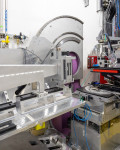
Using the liquids reflectometer neutron instrument at ORNL’s Spallation Neutron Source, the researchers measured and analyzed how neutrons were scattered, or “reflected,” at different angles depending on the composition of the lipid layers they passed through.
Pursuing another angle
“Neutron reflectometry gave us information on the internal structure of the membranes, but we also needed to understand how the outer surfaces of the membranes behave,” said John Katsaras, a biophysicist specializing in neutron scattering and the study of biological membranes at ORNL’s Neutron Sciences Directorate. “It’s like a sheet of paper where we had looked inside the sheet, but we also needed to examine the front and back sides of the paper. So we conducted small-angle neutron scattering experiments, which revealed details about the surfaces of materials.”
The different types of neutron experiments revealed how the membranes reacted to various pressures and tensions while compressed or decompressed, which enabled the scientists to develop a baseline model of how the bare membrane behaves internally and across its surfaces, in a range of conditions and independent of any complicating factors. Supporting experiments were performed at Virginia Tech by Assistant Professor Rana Ashkar, who carried out complementary microscopy studies on the lipid monolayers.
After conducting control experiments, the team inserted E-proteins into the membranes (the lipid models) and examined the structures. They were looking specifically at how the protein’s ion channel and PDZ-binding motif work together and how the ion channel facilitates the virus inserting its genetic material into a cell.
“It’s important that we understand how the ion channel works in this virus, because certain molecules in existing drugs have been shown to be capable of inhibiting E-proteins,” said Jesse Labbé, a cellular and molecular fungal geneticist in the ORNL Biosciences Division. “In laboratories, ion channel blockers have proven effective in limiting inflammation caused by a similar coronavirus, SARS-CoV, which caused a deadly pandemic in 2002. That’s what makes this line of research so promising.”
The structure and activity of the E-protein were further studied by Pat Collier, a cleanroom process engineer from ORNL’s Center for Nanophase Materials Sciences, who examined droplet interface bilayers to record ion channel activity, and by Ben Doughty, a research scientist from ORNL’s Chemical Sciences Division, who conducted vibrational spectroscopy neutron experiments to probe the E-protein’s orientations and secondary structures.
Masters of disguise
Another reason that coronaviruses are so difficult to deal with is that they are masters of disguise, regularly mutating to avoid being recognized by a person’s immune system.
To address the challenges posed by mutations, the scientists at ORNL are focusing on features of the current coronavirus that tend to mutate less frequently from generation to generation. Such “conserved” features, if targeted successfully by a drug compound, would enable developing a treatment with longer lasting effects.
“The team here at ORNL has studied membranes for decades and has a lot of expertise in this area, and membrane function is a key component in COVID-19 disease,” said Jacob Kinnun, postdoctoral researcher with the SNS Reflectometry team. “If our research can identify a mechanism that inhibits the SARS-CoV-2 virus’s ability to trigger inflammation in people infected with COVID-19, it could lead to a treatment that reduces the number of people who need hospitalization and help save lives.”
The researchers next plan to expose the E-proteins to some of the most promising drug compounds that have shown to inhibit similar proteins. One goal is to develop a model of how the SARS-CoV-2 E-protein functions and what drug compounds are most effective against it, enabling researchers worldwide to quickly and accurately screen new drugs to help determine how they might work against future virus outbreaks.
The work was funded by the DOE Office of Science through the National Virtual Biotechnology Laboratory program, a consortium of DOE national laboratories focused on response to COVID-19, with funding provided by the Coronavirus CARES Act; and the ORNL Laboratory Directed Research and Development program. The researchers used resources of the ORNL Center for Structural Molecular Biology, which is supported by the DOE Office of Science, and performed work at ORNL’s Center for Nanophase Materials Sciences.
SNS and CNMS are DOE Office of Science User Facilities. ORNL is managed by UT-Battelle LLC for DOE’s Office of Science, the single largest supporter of basic research in the physical sciences in the United States. DOE’s Office of Science is working to address some of the most pressing challenges of our time. For more information, visit www.energy.gov/science. –by Paul Boisvert