

Contacts
-

Instrument Scientist
-

Instrument Scientist
-

Instrument Scientist
Biochemistry, biotechnology and drug design at the atomic scale
Neutron macromolecular crystallography (NMX) provides unparalleled insight into the molecular mechanisms of biological macromolecules, resolving fundamental questions about enzyme catalysis and enabling new strategies for rational drug design. As the only experimental technique capable of directly determining the positions of hydrogen atoms in biomacromolecules, NMX reveals critical details of hydrogen bonding, protonation states, and proton transfer that underpin biochemical reactions and molecular recognition. With its high-resolution capabilities, IMAGINE-X will enable researchers to investigate proteins, nucleic acids, and their complexes at an unprecedented level of functional detail. These studies will elucidate enzyme mechanisms, guide the engineering of improved industrial enzymes, reveal the molecular basis of disease, and accelerate the development of next-generation therapeutics, vaccines, and other structure-based drug design strategies.
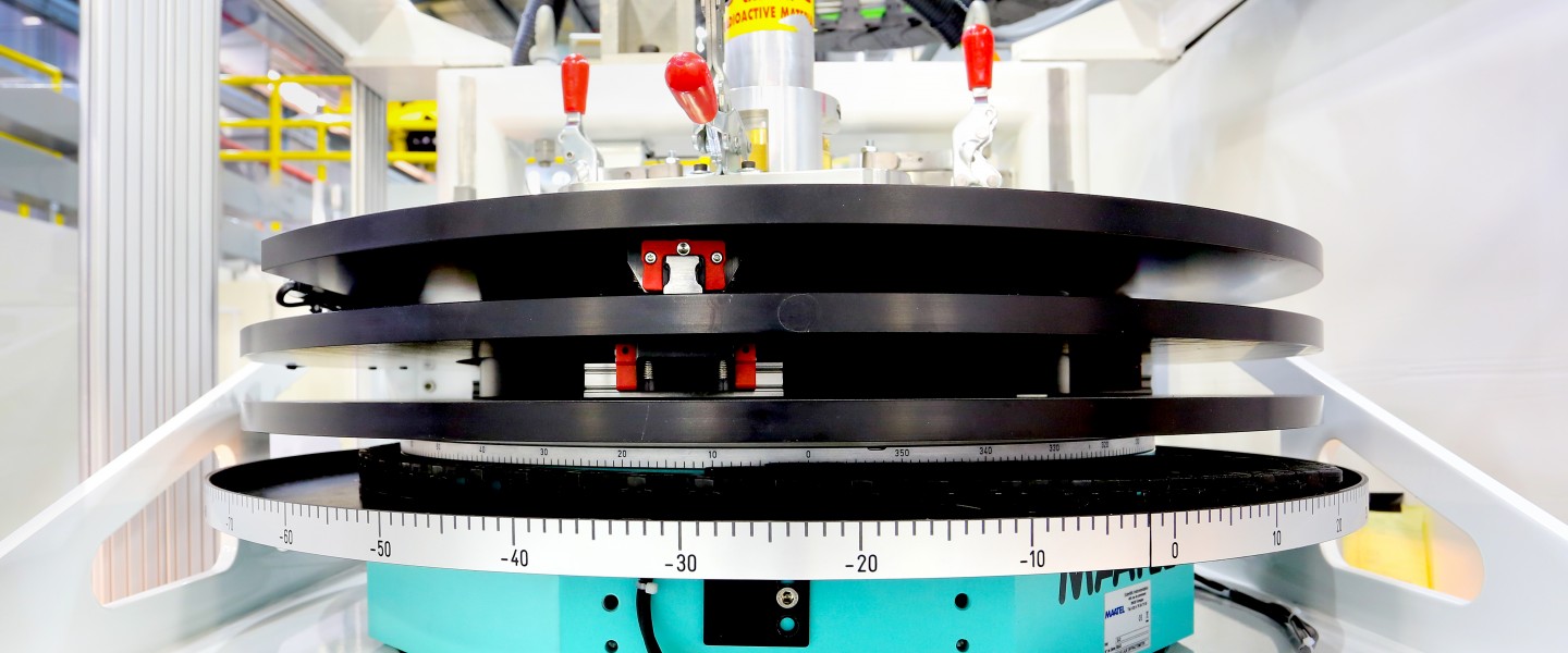
CG-4D’s IMAGINE-X instrument is a state-of-the-art single-crystal diffractometer that provides atomic resolution information on inorganic, organic, metallo-organic, and macromolecular single crystals that enables their chemical, physical, and biological structure and function to be understood. IMAGINE-X benefits communities with interest in pharmaceuticals, minerals and materials, small molecules, molecular organo-metallic complexes and metal-organic frameworks and enables the neutron crystal structure of oligonucleotides and proteins to be determined at near atomic resolutions (1.5 Å).
The instrument has undergone significant upgrade with the design and installation of a new continuously operating dynamic nuclear polarization (DNP) system for nuclear polarization, a new suite of 74 silicon photo multiplier (SiPM) Anger camera detectors suite and new polarized optics and spin-flipper to control the spin state of each event. The new DNP system enables tunable control of hydrogen neutron scattering cross sections in situ within biological crystals during experiments, which will provide order of magnitude enhancements in data collection and in the visibility of hydrogen atoms in the resulting protein structures. The polarized incident beam option also provides enhanced sensitivity for probing weak magnetic scattering signals from magnetic materials.
Macromolecular structure and function
Supra-Molecular Crystallography
Sample Environment: Materials under Extreme Environment
| Flux | ~107 n/s/cm2 |
| Cross section | 2.0 x 3.2 mm |
| Wavelengths minimum | 2.0, 2.8, 3.3 Å |
| Wavelengths maximum | 3.0, 4.0, 4.5 Å |
| Detector | 74 silicon photo multiplier (SiPM) Anger cameras |
| Detector size | 2pi opening angle |
| Pixel size | 500 µm |
| Sample-to-detector distance | Variable – 350-600 mm |
| Goniometer | Multi-axis Kappa, Phi, Omega cryo-goniometer operating at 1K |
| The calculator assumes that only one crystal grows in the drop. Protein solubility is not considered. Accuracy of protein concentration is essential. |
| O’Dell W.B., Bodenheimer A., Meilleur F. (2016) Neutron protein crystallography: A complementary tool for locating hydrogens in proteins. Arch Biochem. Biophys. 602:48-60 |
Instrument Scientist
Instrument Scientist
Instrument Scientist







Oak Ridge National Laboratory is managed by UT-Battelle LLC for the US Department of Energy
