Today’s range of techniques for detection of breast and other cancers include mammography, computed tomography (CT), magnetic resonance imaging (MRI), ultrasound, positron emission tomography (PET), and optical imaging. Each technology has advantages and disadvantages, with limitations either in spatial resolution or sensitivity. Researchers from the University of Tennessee College of Veterinary Medicine in Knoxville (UTCVM) and Oak Ridge National Laboratory (ORNL) are exploring the potential capabilities of imaging with neutrons to aid in detecting breast cancer.
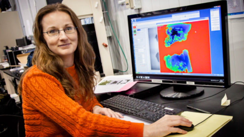
Maria Cekanova, a research assistant professor at the UTCVM Department of Small Animal Clinical Science, is using the hydrogen-sensitive neutron imaging capabilities at the High Flux Isotope Reactor (HFIR) to image healthy and cancerous breast tissue specimens. Working with Hassina Bilheux, lead neutron imaging instrument scientist at ORNL, and collaborators Jean-Christophe Bilheux (ORNL) and Alfred Legendre and Robert Donnell (UTCVM), the team is assessing the value of neutron imaging in examining larger sections of biopsied tissues for diagnosis.
“Several imaging techniques are used for detection and diagnosis of cancer and for monitoring the cancer’s responses to treatment,” says Cekanova. “Current research is focused on early detection of changes during transformation of the tumor in order to use more effective therapies at early stages to prevent progression of the cancer. Our research interest is in the evaluation of biospecimens using neutron radiography and neutron computed tomography to assist pathologists with diagnosis. The advantage of neutron imaging is to obtain the three-dimensional image of a tissue sample without slicing it, as is commonly done during pathology evaluation.”
The breast tissue samples used in the study come from dogs that are patients at UTCVM, where they are being diagnosed and treated for cancers. “We have access to non-utilized tumor tissues after diagnosis is performed at UTCVM,” Cekanova says. “These are valuable samples because tissues obtained from dogs diagnosed with naturally occurring cancers are similar to human cancers.”
“Development of multi-modality imaging technologies for detection of cancer is ongoing,” says Cekanova, who has made cancer research her career. “Neutrons are seen as a potentially powerful complementary imaging technique because they predominantly detect hydrogen in tissue, which is one of the most abundant elements in nature. This work with neutrons could advance detection of not only breast cancer but also of other types of cancer.”
The work is being conducted at the neutron imaging facility (CG-1D) at HFIR. “The reactor at HFIR gives us a broad range of neutron energies and an established way to do neutron imaging,” Bilheux says. “We are very lucky to benefit from HFIR’s intense flux for neutron imaging research.”
“We would like to be able to evaluate a larger block of tissue and get three-dimensional views, not just a two-dimensional thin section,” Cekanova says. “Currently, we can image 2 to 3 mm thick tissues at HFIR because of the neutron energy range (i.e., cold neutrons) available at the facility. Neutron radiography lets us keep the specimen intact. In the future, we will be able to image biopsied tissues of several centimeters thick using the higher-energy neutrons called epithermal and fast neutrons that will hopefully be available soon at the ORNL VENUS instrument (Versatile Neutron Imaging Instrument at SNS).”
Cekanova says that she and Bilheux are excited about the progress they are making at this early stage of their project. “We have our first tumor images and are now analyzing data and learning more about neutron transmission through both the cancerous and normal tissues.”
This research is being funded by the Scientific User Facilities Division, Office of Basic Energy Sciences, US Department of Energy, and the University of Tennessee College of Veterinary Medicine.


