
In the 1950s at Minimata, Japan, 900 people died and 2 million suffered life-long injury, in the form of birth defects to incapacitating neurological and muscular deformities, after swimming in and consuming fish from the bay nearby.
The disaster was caused by the dumping into the bay of mercury compounds, including methylmercury. When ingested, this lethal toxin crosses the blood/brain barrier, with catastrophic results.
Not all mercury is as lethal as is the killer methylmercury. And there is a broad federal effort that includes the U.S. Department of Energy (DOE) and Oak Ridge National Laboratory (ORNL) to understand the chemistry of mercury poisoning and to clean up mercury-polluted sites.
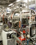
Researchers at the Spallation Neutron Source (SNS) and the ORNL Environmental Sciences Division (ESD) are using the ultrahigh-resolution Neutron Spin Echo (NSE) instrument to study the internal dynamics of a remarkable family of bacteria that eats its way into mercury in nature, transforming it into less toxic forms, without itself being killed in the process. NSE was built by and is operated by FZ Jülich in Germany.
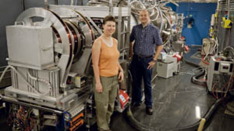
Alongside a large collaboration of researchers, Alex Johs, a young biophysicist, and Melissa Sharp, a young instrument scientist for NSE, are studying this bacterium that in nature sniffs out the presence of toxic mercury, latches onto it, and breaks it down.
Johs, of ESD, works with group leader Liyuan Liang, who four years ago pulled together divergent lab research efforts into a mercury science focus area for DOE’s Office of Biological and Environmental Research.
“We look at the fundamental molecular mechanisms of these proteins to understand how these bacteria are able to deal with high levels of toxic mercury without getting poisoned and killed in the process,” Johs said.
The bacterium is able to motor through mercury like a mini Pac-Man because it has a set of genes that is regulated by a protein called MerR that looks like a metal staple and acts like a traffic cop. This regulator/cop first enables the mercury processing bacteria to detect traces of the metal and then switches on genes in the bacterium’s DNA. Those genes are the blueprints for other proteins and enzymes that protect the bacteria from the toxic effects of mercury and can even help remove it from its environment.
With this powerful arsenal of enzymes on hand, these bacteria can break down the highly toxic methylmercury (CH3Hg+) to methane (CH4) and mercuric mercury (Hg2+), which is subsequently turned into relatively harmless mercury vapor (Hg0).
“The reason why we like to have these bacteria around is because they have the potential to remove significant amounts of methylmercury from the ecosystem that would otherwise enter the food chain and accumulate in higher organisms such as fish,” said Johs. What the researchers are looking at is the special dynamics inside the regulator (MerR) protein that allows the bacteria to detect mercury and initiate the production of the molecular defense machinery that enables it to break down the contaminant.”
Enter the NSE instrument at SNS. The protein is prepared in a buffer solution and then placed at 10°C inside a quartz cell. The cell is then placed in the neutron beam, with a superconducting magnet placed before and after the sample.
The instrument makes use of a neutron property called “spin” to label the neutrons, indicating their velocity. By changing the magnetic field in the superconducting magnets, researchers can measure any change in the neutron velocity caused by the sample, which in turns reveals the motions taking place in the sample. The researchers focus on measuring the motions of the protein on different length scales.
“The measuring times are quite long on spin echo, so we must work to preserve our sample from degradation,” said Sharp. “It is quite complicated for we must subtract the bigger diffusive motions that the protein has from the dynamics inside the protein itself.”
The measurement requires what neutron scientists call a wide Q range. Q is the momentum transfer (kick) the neutron gives to the sample when it scatters off it and is related to the lengthscales probed in the sample.
The Q range indicates the distances over which the dynamics are taking place. A larger Q means a harder kick, which is used to see smaller distances. A small Q sees larger structures. It’s like changing the magnification scale on a microscope.
The team measures a wide time range, from 100 picoseconds (trillionths of a second) to 100 nanoseconds (billionths). “The combination of the different length scales and those time scales enable us to see the protein’s internal motions, the domain motions,” Sharp explained.
Anne O. Summers, a University of Georgia microbiologist who has been studying mercury-resistant bacteria for almost 30 years, is the main external collaborator.
When the experiment is completed, University of Tennessee/ORNL Governor's Chair Jeremy C. Smith will provide computational simulations of the experimental data.
“The supercomputer will be used for the simulation of slow, large-scale molecular motions to interpret the data collected on the NSE instrument. The simulation of very long timescales requires a lot of processing power.”
In recent work, Smith and his computational colleagues have been interpreting data from the NSE instrument, simulating the slow, large-amplitude motions inside biological macromolecules. They identified types of motion that dominate the spin echo signal in the enzyme phosphoglycerate kinase.
“This work demonstrates that the combination of NSE with molecular dynamics code is useful for determining the nature of functional domain motions inside proteins,” said Smith. “It may presage wide application of these techniques in interpreting large-scale motions in biomolecules.”
Collaborating with Sharp [European Spallation Source (ESS AB), Sweden, and the Jülich Centre for Neutron Science, SNS Outstation, USA] and Johs (ORNL) are S. Tomanicek, Z. Yi, H. B. Guo, J. Parks, L. Liang, and J. C. Smith (ORNL), M. Ohl (Jülich Centre for Neutron Science, SNS Outstation, USA); R. Biehl, M. Monkenbusch, and D. Richter (Jülich Centre for Neutron Science & Institute for Complex Systems, Germany); and A. Summers and L. Oliff (University of Georgia).
Research conducted at the Spallation Neutron Source was funded by the U.S. Department of Energy Office of Basic Energy Sciences.